For more information and appointment
For more information and appointment
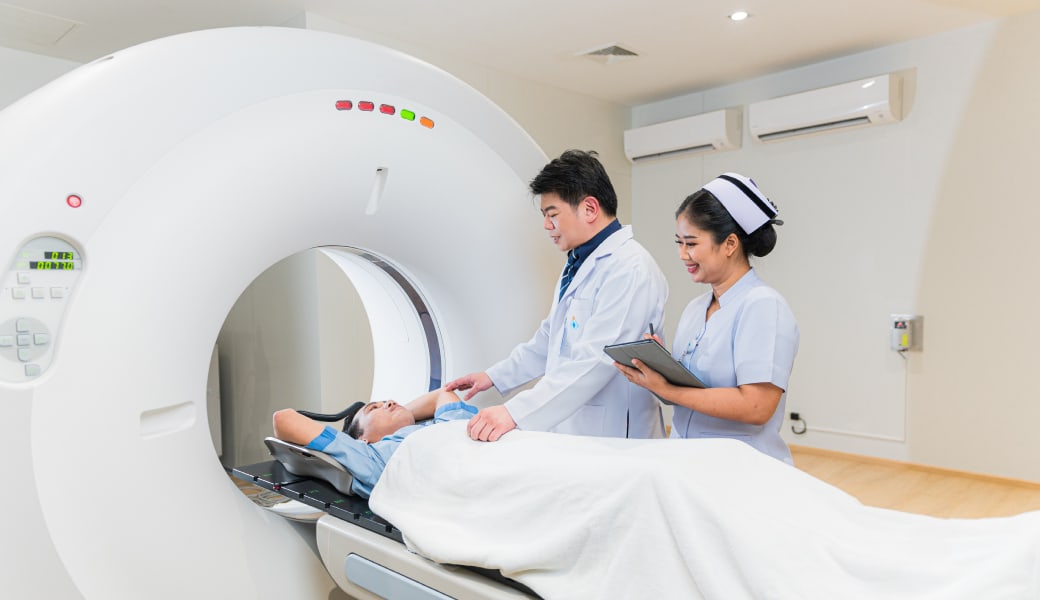
A cancer treatment with radiotherapy. Determining the exact locations of irradiation is extremely important. The Computed Tomography Simulator, or CT Simulator, is a computerized x-ray machine that specifies the position, shape and size of the tumor tissue as well as nearby organs by creating 3D images, allowing physicians to plan the treatment using radiation in multiple directions. In this way, the radiation dose will be distributed consistently and will also reduces the amount of radiation affecting nearby organs. Therefore this method is suitable for complicated lesions.
Chiwamitra Hospital chooses Canon AquilionTM LB for computerized x-ray treatment simulation. A modern computerized x-ray machine with the following special features:
1. It has the widest radiation area of 70 cm., and can be expanded to 85 cm., supporting all organs, every lesion and area. The process all covered in one irradiation, therefore helping to simulate images for diagnosis, and make the treatment plan more accurate.
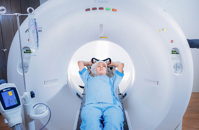
2. The machine cavity is larger than general computer tomography simulators. It has a wide size diameter of 90 cm., making it possible to insert different types of clamp (for immobilization) to allow the patient to remain still during each irradiation and more convenience treatment.
3. The Pure Vision Detector technology has the ability to collect detailed simulation, and able to store images of material as thin as 0.5 mm. The Double Reconstruction technology creates a high image resolution to help plan the best treatment.
4. The Sure Exposure and AIDR3D technology ensure every patients will receive the least amount of radiation from treatment.
5. The Ultra Fast Data Transfer technology has the ability to simulate 22 images per second, allowing faster treatment planning results.
1. No water and food before the examination for 6 - 8 hours.
2. In the following cases, you must inform staff at the time of your appointment or before the examination:
- Women who are pregnant or suspect being pregnant
- Patients who have a history of allergies to radiocontrast agents before
- Patients who have a history of seafood allergies
- Patients who have a history of hypersensitivity to other drugs.
- Patients who have a history of chronic diseases such as allergies, asthma and epilepsy, etc.
3. In case of patients examining the lower and all abdominal systems, they must drink water containing radiocontrast agents according to instructions.
4. Patients who have a history of allergies to radiocontrast agents, seafood, or asthma. Take oral medication or injection as ordered by the doctor.
5. Patients aged over 60 years or having any chronic illnesses should have a kidney function test not more than 7 days before the examination (blood can be checked at the nearest medical facility and results taken on the examination day).
6. All patients must refrain from all food and water. You can take your medicine and drink water, but not more than a 1/4 of a glass of water, two hours before the examination.
1. Allergic reactions may occur after exposure to radiocontrast agents. This can occur immediately after and no more than 7 days after the examination. Symptoms include congestion, itching, swollen eyelids, swollen lips, and swollen larynx. Most allergies are mild allergies, such as blisters, and itching occurs in about 1.4% of people. In some cases, severe symptoms such as low blood pressure, unconsciousness, and cardiac arrest occur, but this is very rare - only 0.04% (4 out of 10,000 people).
2. Side effects from the examination include nausea, vomiting, headache, and muscle pain. Often, these are mild symptoms and disappear by themselves.
3. Adverse reactions to the kidneys may occur but are very rare
4. All abdominal computer tomography patients must drink water or water mixed with radiocontrast agents or take an enema containing these substances. After the examination, nausea, vomiting, and diarrhea may occur. These symptoms will improve and disappear within 2-3 days.